Pick up and Drop
Service Available

Breast health is an essential part of overall well-being, yet many women postpone breast examinations until they notice discomfort or a visible change. While self-examinations are valuable, they cannot reveal everything happening beneath the surface. This is where Breast Imaging becomes an important part of preventive healthcare. Modern imaging techniques allow specialists to detect subtle changes in breast tissue that may not be felt during a physical examination, helping doctors understand the cause of symptoms or monitor breast health more effectively.
Unlike a routine examination, a breast imaging test provides detailed visual information about the internal structure of the breast. It helps healthcare professionals distinguish between normal tissue, fluid-filled cysts, and solid abnormalities that may require additional evaluation. Whether performed as part of routine screening or after symptoms appear, diagnostic breast imaging offers valuable clinical information that supports accurate medical decisions while reducing uncertainty for patients.
Many people associate imaging only with detecting serious diseases, but its purpose extends much further. A breast imaging scan helps evaluate a wide range of breast conditions, from harmless cysts and hormonal changes to suspicious lesions requiring closer examination. By identifying the exact nature of an abnormality, imaging often prevents unnecessary invasive procedures and guides the next steps in patient care.
Different imaging methods are selected based on age, breast density, symptoms, and medical history. Younger women often benefit from a breast ultrasound test, while other patients may require additional imaging depending on clinical findings. Rather than relying on assumptions, imaging provides evidence that helps healthcare providers recommend the most appropriate course of action.
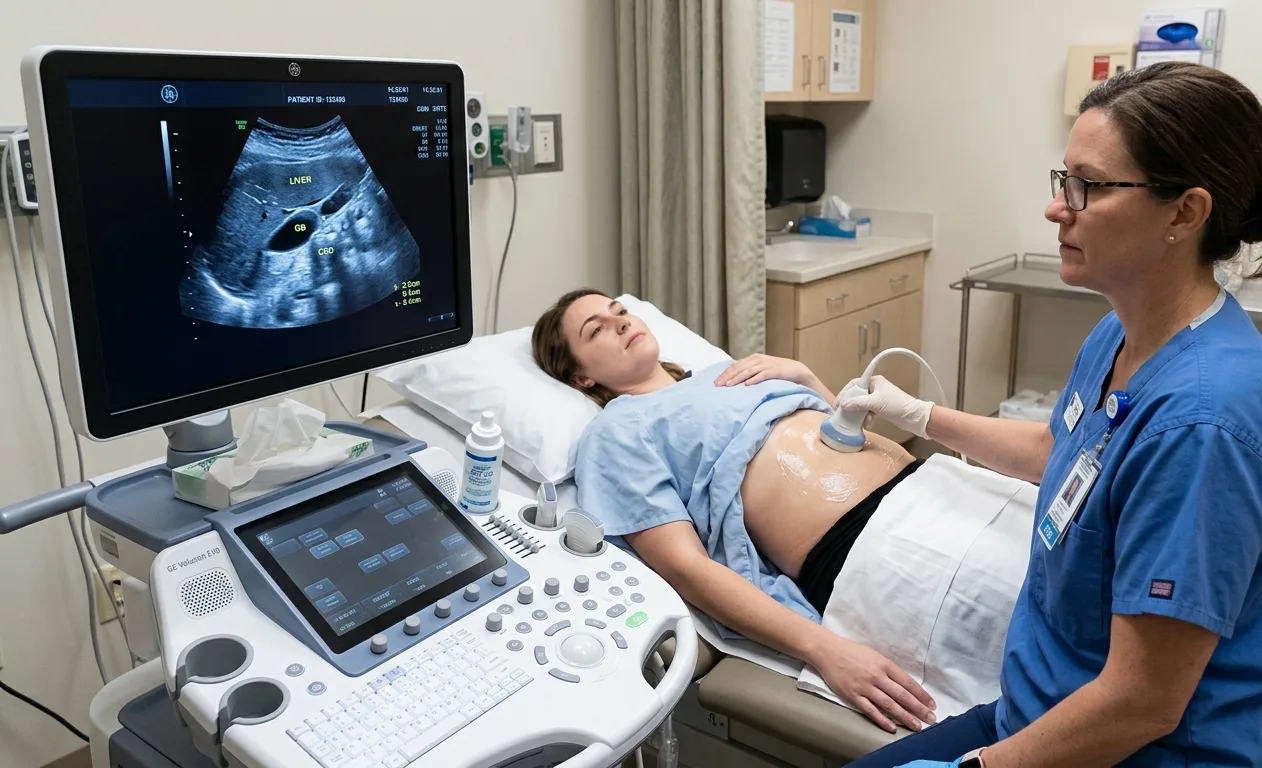
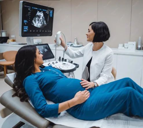
A breast ultrasound test uses high-frequency sound waves to create real-time images of breast tissue without exposing the patient to radiation. This makes it a preferred imaging method for evaluating palpable lumps, breast pain, nipple discharge, and dense breast tissue.
A breast ultrasound scan can help identify:
Because ultrasound differentiates between fluid and solid structures, it is particularly useful for confirming whether a lump is a simple cyst or something that requires additional evaluation.
Many people assume that Breast Imaging is only necessary when symptoms develop, but it also plays a vital role in preventive healthcare. A doctor may recommend a breast imaging test if you notice a lump, persistent breast pain, skin thickening, nipple discharge, or changes in breast size or shape. Women with dense breast tissue, a family history of breast conditions, or previous breast abnormalities may also benefit from regular imaging as advised by their healthcare provider.
A breast ultrasound test is particularly useful for younger women and for evaluating breast changes that require closer examination. Rather than replacing other imaging methods, ultrasound often complements them by providing additional information about the structure and nature of breast tissue. Early evaluation allows healthcare professionals to make informed decisions while reducing unnecessary uncertainty.

Modern Breast Imaging offers several important advantages for both patients and healthcare providers.
These benefits make breast imaging an essential component of comprehensive breast healthcare.
A mammary ultrasound, also known as sonomammography, is a highly effective imaging technique for evaluating breast tissue, especially in women with dense breasts. It produces detailed images without radiation and is often recommended alongside clinical examinations or other imaging methods.
If you are looking for a trusted Sonomammography Centre in Dehradun, choosing an experienced diagnostic facility is essential. Skilled radiologists, high-resolution ultrasound systems, and standardized reporting contribute to more reliable results and better clinical decisions. A detailed breast ultrasound scan helps evaluate cysts, solid masses, and other breast abnormalities with greater confidence.
One of the biggest misconceptions is that Breast Imaging exists only to detect breast cancer. In reality, imaging helps identify a wide range of breast conditions, many of which are completely non-cancerous.
A comprehensive breast ultrasound test can reveal:
Every finding tells a different story. Imaging helps healthcare providers understand whether a breast change is likely to be harmless or whether further clinical investigation is necessary.
Breast conditions can develop gradually without causing noticeable symptoms. By the time physical changes become visible, further evaluation may already be necessary. Diagnostic breast imaging helps identify tissue changes at an earlier stage, making it easier to understand whether a finding is harmless or requires medical attention.
Early assessment is not only about detecting serious disease. Imaging also helps identify benign cysts, fatty tissue changes, hormonal variations, and inflammatory conditions that can cause discomfort. Timely breast lesion evaluation enables doctors to recommend appropriate follow-up while avoiding unnecessary procedures whenever possible.
Choosing the right diagnostic centre is just as important as choosing the right imaging test. At Semwal Diagnostics Centre, every patient receives personalized attention supported by modern imaging technology and experienced radiologists. The centre focuses on delivering detailed and reliable Breast Imaging services while ensuring patient comfort throughout the examination process.
Whether you require a breast ultrasound test, diagnostic breast imaging, breast imaging scan, or detailed breast lesion evaluation, the team follows standardized imaging protocols to provide clear and clinically meaningful reports. Patients also benefit from timely reporting, professional guidance, and a commitment to quality healthcare.
For individuals seeking a dependable Sonomammography Centre in Dehradun, Semwal Diagnostics Centre offers comprehensive breast imaging services designed to support accurate diagnosis and informed treatment decisions.